Category:Acetabulum
Idi na navigaciju
Idi na pretragu
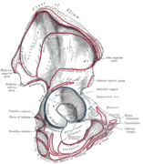
a cavity in the coxal bone where thigh bone (femur) articulate with pelvis | |||||
| Postavi datoteku | |||||
| Je |
| ||||
|---|---|---|---|---|---|
| Je podklasa od |
| ||||
| Je dio | |||||
| Connects with |
| ||||
| Sastoji se od |
| ||||
| Različito od | |||||
| |||||
Potkategorije
Prikazano je 5 potkategorija, od ukupno 5.
A
- Acetabular fossa (24 F)
- Acetabular margin (30 F)
- Acetabular notch (20 F)
L
- Lunate surface of acetabulum (15 F)
Datoteke u kategoriji "Acetabulum"
Prikazane su 52 datoteke u ovoj kategoriji, od ukupno 52.
-
Acet anatomy bone model obturator view3.jpg 240 × 365; 18 KB
-
Acet anatomy bone model1.jpg 240 × 365; 16 KB
-
Acet anatomy bone model2.jpg 246 × 371; 20 KB
-
Acet Ant wall Cooper 01.jpg 1.191 × 1.525; 121 KB
-
Acet Both column 03.jpg 602 × 635; 225 KB
-
Acet tshape function1.jpg 194 × 255; 11 KB
-
Acet tshape02.jpg 226 × 319; 13 KB
-
Acetabolo femore adduzione abduzione.gif 190 × 193; 183 KB
-
Acetabular Labrum Exercises.jpg 631 × 441; 59 KB
-
Acetabulum (model).jpg 330 × 310; 10 KB
-
Acetabulum 00 animation.gif 360 × 360; 1,29 MB
-
Acetabulum 01 animation.gif 360 × 360; 2,52 MB
-
Acetabulum 02 animation.gif 360 × 360; 3,55 MB
-
Acetabulum 03 animation.gif 360 × 360; 2,34 MB
-
Acetabulum 03 anterior view.png 1.125 × 1.125; 306 KB
-
Acetabulum 03 inferior view.png 1.125 × 1.125; 272 KB
-
Acetabulum 04 animation (Right hip bone).gif 360 × 360; 2 MB
-
Acetabulum 04 lateral view (Right hip bone).png 1.125 × 1.125; 207 KB
-
Acetabulum.jpg 960 × 720; 96 KB
-
Biomechanik-Hueftdysplasie.png 640 × 486; 26 KB
-
Biomechanik-Hüftdysplasie-dysplastisch.svg 322 × 338; 5 KB
-
Biomechanik-Hüftdysplasie-Normalstellung.svg 322 × 338; 5 KB
-
Coxal bone - lateral view.jpg 960 × 720; 118 KB
-
Gray235-ar.png 793 × 911; 533 KB
-
Gray235.png 793 × 911; 122 KB
-
Gray237-ar.png 430 × 500; 175 KB
-
Gray237.png 459 × 533; 33 KB
-
Gray321-ar.png 600 × 458; 220 KB
-
Gray321.png 600 × 458; 50 KB
-
Gray321mod.png 1.070 × 908; 234 KB
-
Gray341 zh.png 512 × 500; 157 KB
-
Gray341-ar.png 516 × 500; 275 KB
-
Gray341.png 512 × 500; 47 KB
-
Gray342.png 420 × 450; 32 KB
-
Gray343.png 444 × 500; 48 KB
-
Gray344.png 564 × 500; 225 KB
-
Os coxal face externe.png 935 × 911; 488 KB
-
Os coxal, vue externe, simplifié.jpg 1.389 × 1.809; 712 KB
-
Pelvis - acetabulum (os coxae).jpg 4.608 × 3.456; 5 MB
-
Pelvis - os ischii, acetabulum (lateral).jpg 4.608 × 3.456; 5,2 MB
-
Slide2DAD.JPG 960 × 720; 96 KB
-
Slide2DADA.JPG 960 × 720; 80 KB
-
Slide3A.JPG 960 × 720; 109 KB
-
Slide9AA.JPG 960 × 720; 91 KB
-
Sobo 1909 132.png 1.917 × 1.809; 9,94 MB
-
Sobo 1909 134.png 1.524 × 1.869; 8,16 MB
-
Sobo 1909 136.png 1.380 × 1.659; 1,33 MB
-
Sobo 1909 214.png 1.616 × 1.312; 6,08 MB
-
Sobo 1909 215.png 2.456 × 1.696; 11,94 MB
-
Sobo 1909 216.png 2.404 × 1.680; 11,58 MB
-
Sobo 1909 293.png 996 × 1.484; 4,24 MB
-
Socket - Bone Joints (PSF).png 1.748 × 1.415; 273 KB


