Vesicouterine pouch: Difference between revisions
m Typo/general fixes, replaced: rather then → rather than |
Citation bot (talk | contribs) Add: pmid, authors 1-1. Removed parameters. Some additions/deletions were parameter name changes. | Use this bot. Report bugs. | Suggested by Abductive | Category:Wikipedia articles incorporating text from the 20th edition of Gray's Anatomy (1918) | via #UCB_Category 1769/1775 |
||
| Line 11: | Line 11: | ||
== Structure == |
== Structure == |
||
The vesico-uterine pouch is a fold of [[peritoneum]] over the [[uterus]] and the [[urinary bladder]], forming a pelvic recess.<ref name=":1">{{Citation|last=Hughes|first=Tracey|title=CHAPTER 34 - Pelvic anatomy and scanning techniques|date=2011-01-01|url=http://www.sciencedirect.com/science/article/pii/B9780702031311000341|work=Clinical Ultrasound (Third Edition)|pages=645–659|editor-last=Allan|editor-first=Paul L.|place=Edinburgh|publisher=Churchill Livingstone|language=en|isbn=978-0-7020-3131-1|access-date=2021-02-04|editor2-last=Baxter|editor2-first=Grant M.|editor3-last=Weston|editor3-first=Michael J.}}</ref> It is continued over the intestinal surface and fundus of the [[uterus]] onto its vesical surface, which it covers as far as the junction of the body and [[cervix uteri]], and then to the urinary bladder. It is narrowest when the uterus is [[Anatomical terms of location|anteverted]] rather than [[Retroverted uterus|retroverted]].<ref name=":1" /> The deepest point of the vesico-uterine pouch is typically higher than the deepest point of the [[recto-uterine pouch]].<ref name=":2">{{Cite journal| |
The vesico-uterine pouch is a fold of [[peritoneum]] over the [[uterus]] and the [[urinary bladder]], forming a pelvic recess.<ref name=":1">{{Citation|last=Hughes|first=Tracey|title=CHAPTER 34 - Pelvic anatomy and scanning techniques|date=2011-01-01|url=http://www.sciencedirect.com/science/article/pii/B9780702031311000341|work=Clinical Ultrasound (Third Edition)|pages=645–659|editor-last=Allan|editor-first=Paul L.|place=Edinburgh|publisher=Churchill Livingstone|language=en|isbn=978-0-7020-3131-1|access-date=2021-02-04|editor2-last=Baxter|editor2-first=Grant M.|editor3-last=Weston|editor3-first=Michael J.}}</ref> It is continued over the intestinal surface and fundus of the [[uterus]] onto its vesical surface, which it covers as far as the junction of the body and [[cervix uteri]], and then to the urinary bladder. It is narrowest when the uterus is [[Anatomical terms of location|anteverted]] rather than [[Retroverted uterus|retroverted]].<ref name=":1" /> The deepest point of the vesico-uterine pouch is typically higher than the deepest point of the [[recto-uterine pouch]].<ref name=":2">{{Cite journal|last1=Bricou|first1=Alexandre|last2=Batt|first2=Ronald E.|last3=Chapron|first3=Charles|date=2008-06-01|title=Peritoneal fluid flow influences anatomical distribution of endometriotic lesions: Why Sampson seems to be right|url=http://www.sciencedirect.com/science/article/pii/S0301211508000195|journal=European Journal of Obstetrics & Gynecology and Reproductive Biology|language=en|volume=138|issue=2|pages=127–134|doi=10.1016/j.ejogrb.2008.01.014|pmid=18336988|issn=0301-2115}}</ref> |
||
=== Variation === |
=== Variation === |
||
| Line 17: | Line 17: | ||
== Clinical significance == |
== Clinical significance == |
||
The vesico-uterine pouch may become attached to the [[uterus]], preventing sliding of the [[urinary bladder]] past the uterus.<ref name=":0">{{Citation| |
The vesico-uterine pouch may become attached to the [[uterus]], preventing sliding of the [[urinary bladder]] past the uterus.<ref name=":0">{{Citation|last1=Porter|first1=Misty Blanchette|title=Chapter 35 - Pelvic Imaging in Reproductive Endocrinology|date=2019-01-01|url=http://www.sciencedirect.com/science/article/pii/B9780323479127000354|work=Yen and Jaffe's Reproductive Endocrinology (Eighth Edition)|pages=916–961.e5|editor-last=Strauss|editor-first=Jerome F.|place=Philadelphia|publisher=Elsevier|language=en|isbn=978-0-323-47912-7|access-date=2021-02-04|last2=Goldstein|first2=Steven|editor2-last=Barbieri|editor2-first=Robert L.}}</ref> This may occur in a third of women who have had a [[Caesarean section|caesarian section]], and some people with [[endometriosis]].<ref name=":0" /> |
||
Th vesico-uterine pouch is an important anatomical landmark for chronic [[endometriosis]]. Endometrial seeding in this region causes cyclical pain in women of child-bearing age. This pouch is also an important factor in a [[retroverted uterus]], which can frequently complicate [[pregnancies]]. |
Th vesico-uterine pouch is an important anatomical landmark for chronic [[endometriosis]]. Endometrial seeding in this region causes cyclical pain in women of child-bearing age. This pouch is also an important factor in a [[retroverted uterus]], which can frequently complicate [[pregnancies]]. |
||
Revision as of 14:16, 28 February 2021
| Vesico-uterine pouch | |
|---|---|
 Sagittal section of the lower part of a female trunk, right segment. (Excavatio vesicouterina labeled at bottom right.) | |
 The epiploic foramen, greater sac or general cavity (red) and lesser sac, or omental bursa (blue). Uterovesical excavation labeled at bottom left, third from the bottom. | |
| Details | |
| Identifiers | |
| Latin | excavatio vesicouterina |
| TA98 | A10.1.02.504F |
| TA2 | 3724 |
| FMA | 14729 |
| Anatomical terminology | |
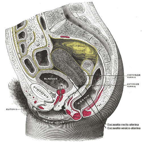
In human female anatomy, the vesico-uterine pouch, also known by various names, is a fold of peritoneum over the uterus and the urinary bladder. Like the recto-uterine pouch, it is a female pelvic recess. However, it is a shallower pouch close to the anterior fornix of the vagina.
Structure
The vesico-uterine pouch is a fold of peritoneum over the uterus and the urinary bladder, forming a pelvic recess.[1] It is continued over the intestinal surface and fundus of the uterus onto its vesical surface, which it covers as far as the junction of the body and cervix uteri, and then to the urinary bladder. It is narrowest when the uterus is anteverted rather than retroverted.[1] The deepest point of the vesico-uterine pouch is typically higher than the deepest point of the recto-uterine pouch.[2]
Variation
When the uterus is very anteverted, the vesico-uterine pouch is deeper than usual.[2]
Clinical significance
The vesico-uterine pouch may become attached to the uterus, preventing sliding of the urinary bladder past the uterus.[3] This may occur in a third of women who have had a caesarian section, and some people with endometriosis.[3]
Th vesico-uterine pouch is an important anatomical landmark for chronic endometriosis. Endometrial seeding in this region causes cyclical pain in women of child-bearing age. This pouch is also an important factor in a retroverted uterus, which can frequently complicate pregnancies.
History
Etymology
The vesico-uterine (or vesicouterine) pouch is also called the vesico-uterine (or vesicouterine) excavation, utero-vesical (or uterovesical) pouch, or excavatio vesicouterina. The combining forms reflect the bladder (vesico-, -vesical) and uterus (utero-, -uterine).
Additional images
-
Median sagittal section of female pelvis.

See also
References
This article incorporates text in the public domain from page 1152 of the 20th edition of Gray's Anatomy (1918)
- ^ a b Hughes, Tracey (2011-01-01), Allan, Paul L.; Baxter, Grant M.; Weston, Michael J. (eds.), "CHAPTER 34 - Pelvic anatomy and scanning techniques", Clinical Ultrasound (Third Edition), Edinburgh: Churchill Livingstone, pp. 645–659, ISBN 978-0-7020-3131-1, retrieved 2021-02-04
- ^ a b Bricou, Alexandre; Batt, Ronald E.; Chapron, Charles (2008-06-01). "Peritoneal fluid flow influences anatomical distribution of endometriotic lesions: Why Sampson seems to be right". European Journal of Obstetrics & Gynecology and Reproductive Biology. 138 (2): 127–134. doi:10.1016/j.ejogrb.2008.01.014. ISSN 0301-2115. PMID 18336988.
- ^ a b Porter, Misty Blanchette; Goldstein, Steven (2019-01-01), Strauss, Jerome F.; Barbieri, Robert L. (eds.), "Chapter 35 - Pelvic Imaging in Reproductive Endocrinology", Yen and Jaffe's Reproductive Endocrinology (Eighth Edition), Philadelphia: Elsevier, pp. 916–961.e5, ISBN 978-0-323-47912-7, retrieved 2021-02-04
External links
- Anatomy photo:43:02-0102 at the SUNY Downstate Medical Center - "The Female Pelvis: Distribution of the Peritoneum in the Female Pelvis"
- Anatomy image:9612 at the SUNY Downstate Medical Center
- Anatomy image:9736 at the SUNY Downstate Medical Center
- Anatomy image:9758 at the SUNY Downstate Medical Center
- figures/chapter_35/35-8.HTM: Basic Human Anatomy at Dartmouth Medical School
This anatomy article is a stub. You can help Wikipedia by expanding it. |