Geert Litjens
GeertLitjens
- Netherlands
- Radboud University Medical Center
- Pathology
- Website
Organizations
Statistics
- Member for 8 years, 7 months
- 43 challenge submissions
- 43 algorithms run
Activity Overview
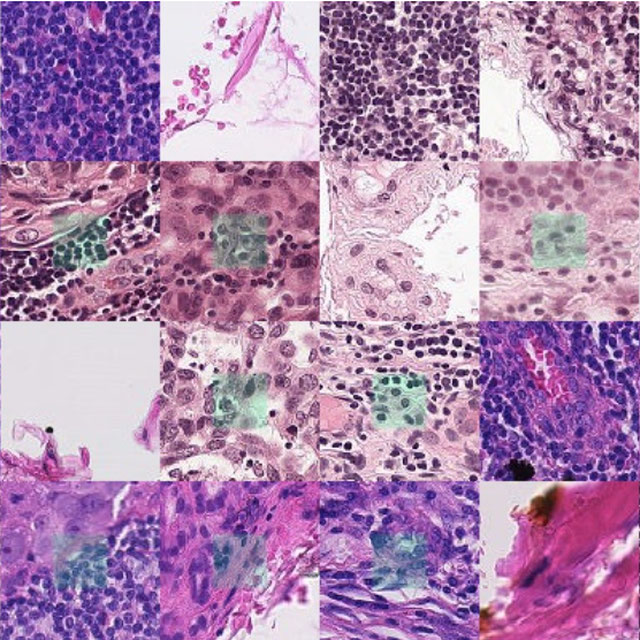
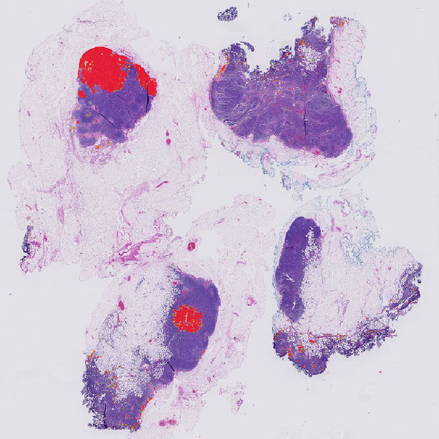
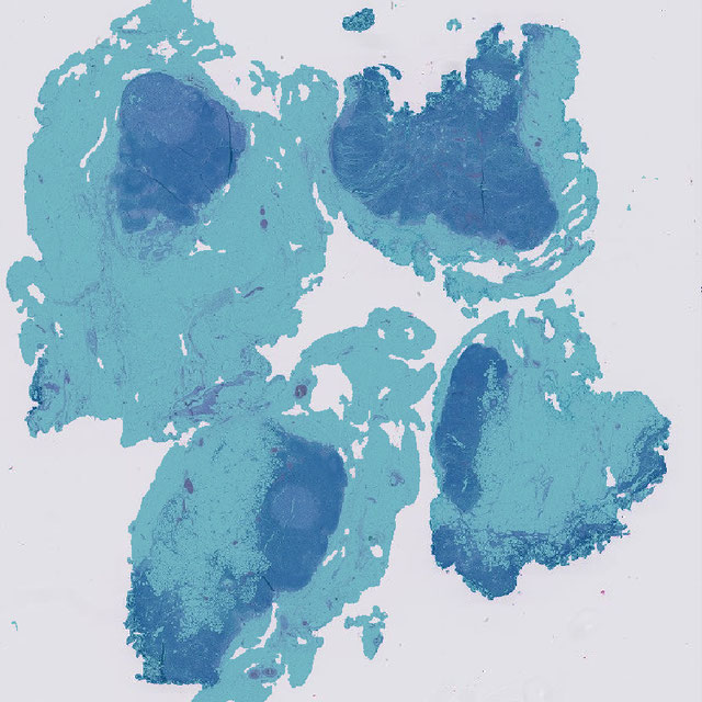
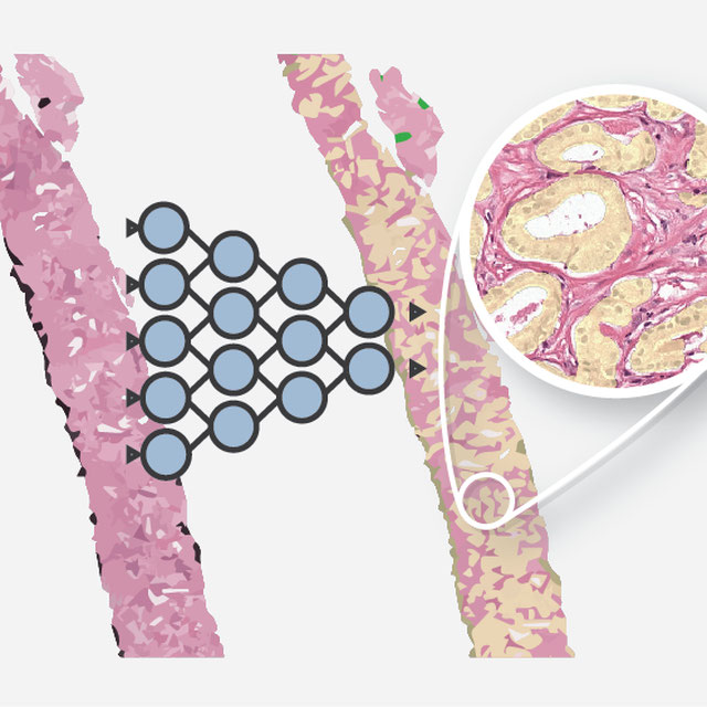
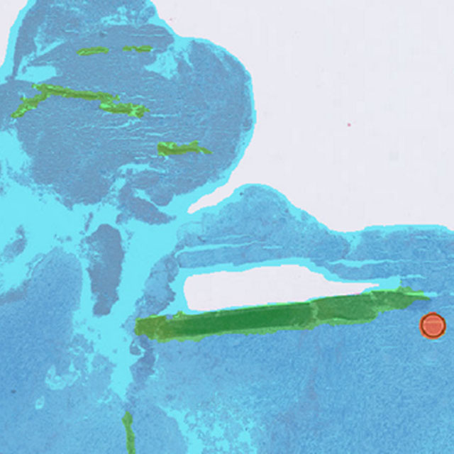
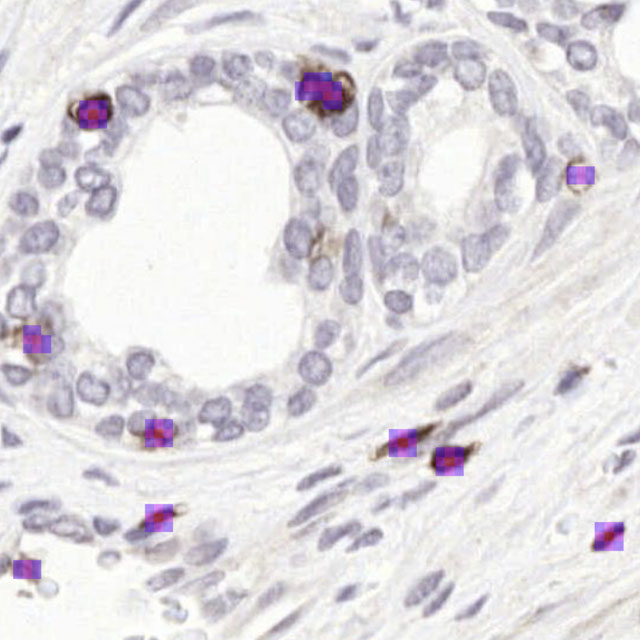
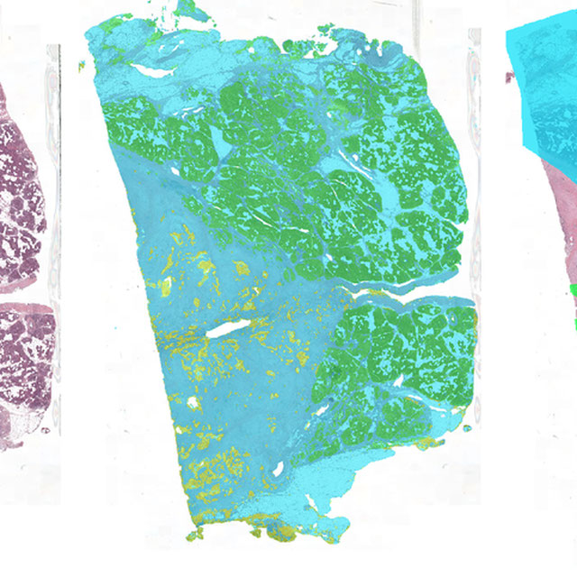
CAMELYON16
Challenge EditorThe goal of this challenge is to evaluate new and existing algorithms for automated detection of cancer metastasis in digitized lymph node tissue sections. Two large datasets from both the Radboud University Medical Center and the University Medical Center Utrecht are provided.