Histology
Histology (from the Greek ἱστός) is the study of tissue sectioned as a thin slice, using a microtome. It can be described as microscopic anatomy. Histology is an essential tool of biology.
Histopathology, the microscopic study of diseased tissue, is an important tool of anatomical pathology since accurate diagnosis of cancer and other diseases usually requires histopathological examination of samples.
The trained scientists who perform the preparation of histological sections are Histotechnicians, Histology Technicians (HT), Histology Technologists (HTL), Medical Scientists, Medical Laboratory Technicians or Biomedical scientists. Their field of study is called histotechnology.
Source of tissue
Histological examination of tissues starts with surgery, biopsy or autopsy.
Technical procedure
Fixation
The tissues are fixed in a fixative, a process that stabilizes the tissues to prevent decay. The most common fixative is neutral buffered formalin (10% formaldehyde in phosphate buffered saline (PBS)).
Embedding
The most common technique is wax embedding. The samples are immersed in multiple baths of progressively more concentrated ethanol to dehydrate the tissue, followed by a clearing agent such as, xylene or Histoclear, and finally hot molten paraffin wax (impregnation). During this 12 to 16 hour process, paraffin wax will replace the water: soft, moist tissues are turned into a hard paraffin block, which is then placed in a mould containing more molten wax (embedded) and allowed to cool and harden.
Embedding can also be accomplished using frozen, non-fixed tissue in a freezing medium. This freezing medium is liquid at room temperature but when cooled will solidify. Non-fixed tissue allows for procedures such as in-situ hybridizations for specific mRNA that would have been destroyed during the fixing process. It also allows for very short turnaround where that is needed, as with a patient currently undergoing surgery.
Sectioning
The tissue is then sectioned into very thin (2 - 8 micrometer) sections using a microtome. These slices, usually thinner than the average cell, are then placed on a glass slide for staining.
Frozen tissue embedded in a freezing medium is cut on a microtome in a cooled machine called a cryostat.
Staining
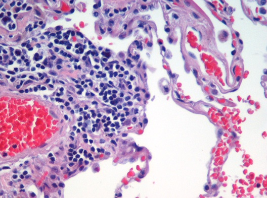
To see the tissue under a microscope, the sections are stained with one or more pigments. This is done to give contrast to the tissue being examined, as without staining it is very difficult to see differences in cell morphology. Hematoxylin and eosin (abbreviated H&E) are the most commonly used stains in histology and histopathology. Hematoxylin colors nuclei blue, eosin colors the cytoplasm pink. There are hundreds of various other techniques which have been used to selectively stain cells and cellular components. Other compounds used to color tissue sections include safranin, oil red o, congo red, fast green FCF, silver salts and numerous natural and artificial dyes, that were usually originated from the development dyes for the textile industry.
Histochemistry refers to the science of using chemical reactions between laboratory chemicals and components within tissue. A commonly performed histochemical technique is the Perls Prussian blue reaction, used to demonstrate iron deposits in diseases like Hemochromatosis.
Recently, antibodies are used to specifically visualise proteins, carbohydrates and lipids: this is called immunohistochemistry. This technique has greatly increased the ability to identify categories of cells under a microscope. Other advanced techniques include in situ hybridization to identify specific DNA or RNA molecules, and confocal microscopy. Digital cameras are increasingly used to capture histological and histopathological images.
Alternative techniques
Alternative techniques include cryosection. The tissue is frozen and cut using a cryostat. They are stained in simular ways to that of wax sections. Plastic embedding is commonly used in the preparation of material for electron microscopy. Tissues are embedded in epoxy resin. Very thin sections (less than 0.1 micrometers) are cut using diamond or glass knives. The sections are stained with electron dense stains (uranium and lead) so that they can be seen with the electron microscope.
History
In the 19th century, histology was an academic discipline in its own right. The 1906 Nobel Prize in Physiology or Medicine was awarded to two histologists, Camillo Golgi and Santiago Ramón y Cajal. They had dueling interpretations of the neural structure of the brain based in differing interpretations of the same images.
Histological classification of animal tissues
There are four basic types of tissues: muscle tissue, nervous tissue, connective tissue, and epithelial tissue. All tissue types are subtypes of these four basic tissue types (for example blood cells are classified as connective tissue since they generally originate inside bone marrow).
- Epithelium: the lining of glands, bowel, skin and some organs like the liver, lung, kidney,
- Endothelium: the lining of blood and lymphatic vessels,
- Mesothelium: the lining of pleural, peritoneal and pericardial spaces,
- Mesenchyme: the cells filling the spaces between the organs, including fat, muscle, bone, cartilage and tendon cells,
- Blood cells: the red and white blood cells, including those found in lymph nodes and spleen,
- Neurons: any of the conducting cells of the nervous system,
- Germ cells: reproductive cells, spermatozoa in men, oocytes in women,
- Placenta: an organ characteristic of true mammals during pregnancy, joining mother and offspring, providing endocrine secretion and selective exchange of soluble, but not particulate, blood borne substances through an apposition of uterine and trophoblastic vascularised parts, and
- Stem cells: cells able to turn into one or several of the above types.
Note that tissues from plant, fungus and microorganisms can also be examined histologically. Their structure is very different from animal tissue.
Related sciences
- Cytology, the study of loose cells, for example cells taken from the cervix during a cervicovaginal smear (pap smear). The cells are directly spread on a glass slide and stained.
- Cell biology is the study of living cells, their DNA, RNA and the proteins they express.
- Anatomy, is the study of organs visible by the naked eye; and
- Morphology, which studies entire organisms.
Histological artifacts
A histological artifact is a structure or feature that is absent in living tissues, but introduced during preparation or staining. Troubleshooting and minimizing artifacts is a major part of the discipline of histochemistry.
References
1. Merck Source (2002). Dorland's Medical Dictionary. Retrieved 2005-01-26.
2. Stedman's Medical Dictionaries (2005). Stedman's Online Medical Dictionary. Retrieved 2005-01-26.
See also
- Pathology
- Anatomical pathology
- Histopathology
- Biological staining
- Important publications in histology